

ARCHIVES
Potential Briefs
ISSUE #04 — July 2011
Comment on this Newsletter... CLICK HERE
ANNOUNCEMENTS
Annual ASET Conference
Stop by the ORIMtec booth #206 and page through our recently released 3rd edition of the Neurotechnologist Reference Guide. See why over 800 copies have been distributed worldwide.
While you are there subscribe to our monthly IONM newsletter Potential Briefs. To stay abreast of the latest in IONM, become a registered member in the IOM Forum.
Watch our Cervical Surgical Procedures training video developed from the popular Surgical Procedures presentation by Dan Slepian, PA, CNIM.
ARTICLE HIGHLIGHT
Dhiraj Jeyanandarajan, M.D. - Board Certified Neurologist
Article: Predictive value of intraoperative neurophyisiological monitoring during cervical spine surgery: a prospective analysis of 1055 consecutive patients
Authors: Michael O. Kelleher, Gamaliel Tan, Roger Sarjeant, Michael Fehlings
Journal: J Neurosurg Spine 8: 215-221, 2008
Summary: This paper is a prospective study of a consecutive series of 1055 patients at an academic centers neurosurgery service who underwent cervical surgery over a 5 year period. The sensitivity, specificity, positive and negative predictive values were determined for somatosensory evoked potentials (SSEP), motor evoked potentials(MEP), and spontaneous electromyography (sEMG).
The author's conclusions are that multimodality monitoring is useful for predicting and preventing neurological injury in cervical spine surgery. SSEPs had a sensitivity or 52%, specificity of 100%, PPV of 100% and NPV of 97%. Motor evoked potential sensitivity was 100%, specificity 96%, and NPV 100%. sEMG had a sensitivity of 46%, speicificity of 73%, PPV of 3% and NPV of 97%.
Comments: Universal acceptance to the benefits of monitoring cervical procedures with intraoperative monitoring is not as prevalent as for thoracolumbar procedures. This paper helps to quantify the utility of the different monitoring modalities and shows...
MORE
TRAINING VIDEO CLIP
Surgical Procedures - Cervical (Disc Level Identification)
ORIMtec Online offers a series of training modules delivered to you via our virtual classroom. Each module is designed to enhance the capabilities and increase the knowledge of the intraoperative neurodiagnostic technologist, guiding him or her to the practical application of electrodiagnostics to a surgical procedure.
View a recorded sample of a live session below. TO REGISTER Click Here!
INSTRUMENT REVIEW
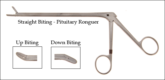
Pituitary Ronguer
The pituitary ronguer is an instrument commonly associated with spinal procedures. This instrument can be used to remove soft tissue, disc material and bone with a biting action. The ronguer can vary in size and the angle of the bite can be straight, up, or down. The tip can be cupped or toothed.
MEP MUSCLEs
Extensor Carpi Radialis Brevis, Vastus Lateralis, Extensor Digitorum Brevis
Transcranial electric motor evoked potentials are frequently requested during cervical surgeries involving the anterior approach, particularly when removing disc material with the pituitary rongure and when impacting the bone graft. The most reliable MEP muscles are those used for fine dexterous movements such as picking up a dime from the table. Three particularly reliable muscles are the abductor pollicis brevis innervated by C8 and T1, the extensor hallucis longus innervated by L5 or flexor digitorum brevis innervated by S1 and S2.
(Bolded is the primary nerve root innervation.)
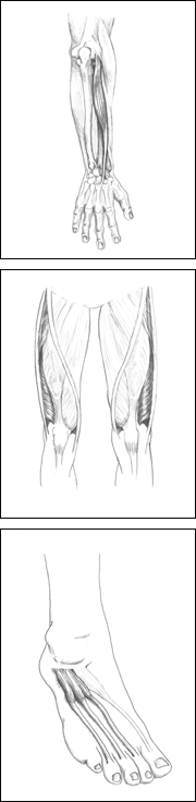 Extensor Carpi Radialis Brevis
Action: Acting with the extensor carpi ulnaris, extends the wrist.
Innervation: .Posterior interosseous nerve, C7 (primary) and C8.
Vastus Lateralis
Action: Extends and rotates the leg, helps to maintain the
patella in its groove during extension.
Innervation: Femoral nerve, L2, L3 (primary), and L4 (primary).
Extensor Digitorum Brevis
Action: Extends the phalanges of the middle three toes.
Innervation: Lateral terminal branch of the deep peroneal
nerve, L5 and S1
Note: This site, along with other muscles of the distal extremities,
has been found to be optimal for tceMEPs. Additional muscle
groups may be monitored to provide added information, but the data is generally considered to be less reliable.